Knee Arthritis (Osteoarthritis)
Knee arthritis is the most common reason for knee replacement surgery in the UK. If knee pain, morning stiffness, or difficulty with stairs is limiting your daily life, Mr Shakir Hussain (Consultant Orthopaedic Surgeon at the Royal Orthopaedic Hospital Birmingham) offers both total and partial knee replacement, including robotic surgery with the MAKO and CORI systems, matched to your anatomy and activity level. With over 5,000 procedures performed and a Doctify rating of 4.98/5, he has the experience to restore your mobility.

What is knee arthritis?
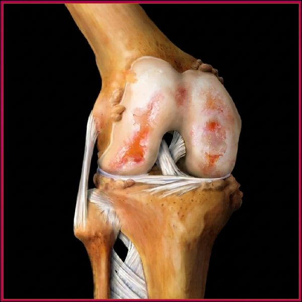
Knee osteoarthritis is the gradual loss of articular cartilage in the knee joint. Without cartilage to cushion the joint surfaces, bone rubs on bone, causing pain, swelling, and progressive stiffness. The medial (inner) compartment of the knee is most commonly affected first, often leading to a bow-legged appearance over time.
The knee is formed by three bones: the femur (thighbone), tibia (shinbone), and patella (kneecap). These surfaces meet across three compartments: medial (inner side), lateral (outer side), and patellofemoral (behind the kneecap). In a healthy knee, smooth articular cartilage coats each surface and allows pain-free movement through a full range of motion.
In knee osteoarthritis, this cartilage thins progressively over years. As it wears away, the underlying bone thickens, bone spurs (osteophytes) form around the joint margins, and the joint lining (synovium) reacts to cartilage debris by producing excess fluid. The knee may develop a bow-legged (varus) or knock-kneed (valgus) deformity as the affected compartment collapses under load.
Knee osteoarthritis is the most common reason for knee replacement surgery in the UK. The National Joint Registry recorded over 100,000 primary knee replacements in 2024, the large majority performed for osteoarthritis.
What does knee arthritis pain feel like?
Knee arthritis typically causes pain on the inner (medial) side of the knee that worsens with walking, standing up from a chair, and descending stairs. Morning stiffness lasting more than 30 minutes is common, along with swelling after activity and a grinding or clicking sensation during movement.
Knee osteoarthritis builds gradually. Symptoms often fluctuate early on, with flares lasting days to weeks followed by periods of relative ease. Over time, pain becomes more persistent, walking distance shortens, and activities that were once routine become increasingly difficult.
Medial knee pain
Deep, aching pain on the inner side of the knee, most noticeable when walking on uneven ground, climbing stairs, or standing up from a low chair or car seat.
Swelling after activity
The knee fills with excess fluid after walking or standing for a prolonged period. Persistent swelling at rest signals more advanced disease and warrants prompt assessment.
Morning stiffness over 30 minutes
Stiffness on waking that lasts more than half an hour before easing. This is notably longer than hip arthritis morning stiffness and is a useful distinguishing feature.
Crepitus (grinding or clicking)
A grinding, creaking, or clicking sensation felt and sometimes heard during knee movement, caused by roughened cartilage surfaces or loose fragments moving inside the joint.
Reduced walking distance
The distance you can walk before pain or stiffness forces a stop shortens steadily over months to years. This is one of the most reliable markers of disease progression.
Pain on stairs and slopes
Descending stairs is often more painful than ascending because the patellofemoral joint (behind the kneecap) bears very high compressive forces when the knee bends on the way down.
What causes knee arthritis?
Knee arthritis develops when cartilage wears down faster than the body can repair it. Key risk factors include age over 50, obesity, previous knee injury, and occupational loading. The link between obesity and knee osteoarthritis is particularly strong: each extra stone of body weight adds roughly four to six times that force through the knee joint with every step.
In primary osteoarthritis, no single cause is identified. It is the cumulative effect of loading on a knee whose repair capacity has declined with age. In secondary osteoarthritis, a previous injury, inflammatory condition, or structural problem is the primary driver.
The most important risk factors are:
- Age over 50. The knee accumulates the highest loads of any lower-limb joint over a lifetime; cartilage repair slows significantly in older tissue.
- Obesity (BMI over 29). Each extra stone of body weight generates four to six times that force through the knee with each step. The relationship between weight and knee arthritis is stronger than for any other joint.
- Previous knee injury. Prior meniscal tears, ACL rupture, or intra-articular fracture permanently alters knee mechanics and accelerates cartilage loss, often by decades.
- Female sex after the menopause. Women have a significantly higher prevalence of knee osteoarthritis than men, partly due to hormonal changes affecting cartilage protection after the menopause.
- Family history. A parent or sibling with knee osteoarthritis increases personal risk by two to three times, reflecting both structural and metabolic inheritance.
- Occupational loading. Years of kneeling, squatting, or heavy lifting in trades such as farming, construction, and floor-laying are associated with earlier onset.
- Quadriceps weakness. Weak thigh muscles reduce the knee's ability to absorb impact under load, increasing cartilage stress. This can be both a cause and a consequence of advancing arthritis.
How is knee arthritis diagnosed?
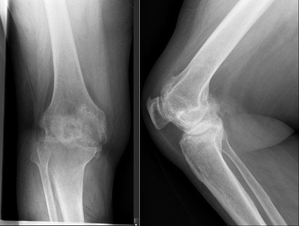
Diagnosis begins with a clinical history and examination. Weight-bearing AP and lateral X-rays of the knee confirm the diagnosis and grade severity using the Kellgren-Lawrence scale from 1 (doubtful) to 4 (severe). Long-leg alignment films are taken before surgery to plan implant positioning accurately.
Mr Hussain will take a careful history covering the location and character of your pain, how far you can walk before stopping, which activities you have given up, how your sleep is affected, and what medications you have tried. Examination assesses the range of knee motion, joint-line tenderness, presence of an effusion, ligament stability, lower-limb alignment, and gait pattern.
Imaging confirms the diagnosis and guides treatment planning:
- Weight-bearing AP knee X-ray. The primary imaging investigation. It shows joint space narrowing (the most important finding), osteophytes at joint margins, subchondral sclerosis, and cysts. Weight-bearing is essential because a non-weight-bearing view can mask significant cartilage loss.
- Lateral knee X-ray. Assesses the patellofemoral compartment and allows measurement of patellar height and the posterior slope of the tibial plateau.
- Kellgren-Lawrence grading. Grade 1 doubtful, Grade 2 mild (definite osteophytes, possible joint-space narrowing), Grade 3 moderate (marked narrowing, sclerosis, possible deformity), Grade 4 severe (gross narrowing, bone-on-bone contact, marked deformity).
- Long-leg alignment film (hip-to-ankle view). Measures the mechanical axis of the leg, essential before knee replacement to plan implant positioning and determine whether deformity correction is required during surgery.
- MRI. Reserved for younger patients where meniscal pathology, ligament injury, or detailed cartilage mapping is needed before deciding between non-surgical management and surgery.
- Blood tests. Only requested if inflammatory or septic arthritis is suspected: morning stiffness lasting over one hour, systemic symptoms, or an atypical age of onset.
Can knee arthritis be treated without surgery?
Yes, for mild to moderate knee arthritis. Structured physiotherapy, weight loss, and anti-inflammatory medication can control symptoms for months to years. NICE recommends therapeutic exercise as the cornerstone of non-surgical treatment. When symptoms substantially affect daily life despite three to six months of conservative care, knee replacement surgery becomes the appropriate next step.
NICE guidance (NG226, updated 2026) recommends a structured non-surgical programme before surgery is considered. Key elements include:
- Therapeutic exercise. Strengthening the quadriceps, hamstrings, and hip abductors reduces knee joint loading and improves stability. This has the strongest evidence base of any intervention for knee osteoarthritis.
- Weight loss. Even a modest 5 per cent reduction in body weight measurably reduces knee pain and load. Weight loss also improves surgical outcomes if replacement becomes necessary.
- Topical NSAIDs. Preferred first-line analgesic, with a good local effect and far fewer systemic side effects than oral tablets. Particularly suited to older patients with kidney or stomach concerns.
- Oral NSAIDs. Used at the lowest effective dose for the shortest duration when topical treatment is insufficient, with attention to cardiovascular and gastrointestinal risk.
- Paracetamol. NICE 2026 no longer routinely recommends paracetamol for osteoarthritis pain, having concluded it is not effective enough on its own to justify routine use.
- Intra-articular corticosteroid injection. Image-guided injection into the knee provides 4 to 12 weeks of pain relief, useful as a bridge before surgery or to facilitate a physiotherapy programme.
- Hyaluronic acid injection. Modest evidence for 4 to 6 months of symptom relief in selected patients, particularly those who cannot tolerate corticosteroids.
- Offloading knee brace. In medial compartment arthritis, a valgus offloading brace shifts load toward the less-affected lateral compartment, reducing pain during walking.
Non-surgical treatment becomes inadequate when arthritis reaches Kellgren-Lawrence grade 3 to 4, when walking distance falls below 100 metres, when night pain disturbs sleep more than twice per week, or when three to six months of structured care has not controlled symptoms. At that point, knee replacement is the right discussion.
If you take prescribed medication, particularly blood-thinners, anti-inflammatories, or rheumatology drugs, please review Mr Hussain's patient guide on medications to pause before hip or knee surgery as you approach a surgical decision.
When should I consider knee replacement?
Knee replacement becomes the right choice when conservative treatment has been tried for three to six months and pain still substantially affects daily life, walking distance is reduced below 100 metres, sleep is disturbed by night pain, or imaging shows end-stage (Kellgren-Lawrence grade 3 to 4) arthritis affecting one or more compartments of the knee.
There is no single test that signals it is time for surgery. The decision is made jointly between you and your surgeon, based on how much the knee is affecting your life. NICE NG226 summarises the threshold as referral for joint replacement when symptoms have a substantial impact on quality of life and non-surgical management is ineffective or unsuitable.
Practical markers Mr Hussain looks for in consultation:
- Pain that wakes you at night, more than twice a week
- Walking distance reduced to under 100 metres before needing to stop
- Significant difficulty getting in and out of a car or rising from a chair
- Pain that substantially limits your ability to manage stairs or slopes
- Three to six months of structured physiotherapy and medication that has not adequately controlled symptoms
- Imaging showing Kellgren-Lawrence Grade 3 or 4 disease
- Withdrawal from activities you previously enjoyed, including walking, travel, and sport
Private consultations with Mr Hussain are available at the Royal Orthopaedic Hospital, Priory Hospital Edgbaston, and Harborne Hospital. See the private consultation and surgery fees page for self-pay pricing and the list of recognised insurers, or book a consultation directly.
Total or partial knee replacement: which is right for me?
Mr Hussain offers both total knee replacement and partial (unicompartmental) knee replacement, with robotic assistance available for both. Total replacement suits patients with arthritis across two or more knee compartments. Partial replacement is best for patients whose arthritis is confined to a single compartment, with intact cruciate ligaments and a BMI under 40.
Both operations relieve the pain of knee arthritis and restore mobility, but they differ in how much of the knee is replaced and which patients benefit most. Mr Hussain is trained in both techniques, certified in robotic-assisted surgery on three platforms (MAKO, ROSA, and CORI), and offers each based on what is right for the individual patient.
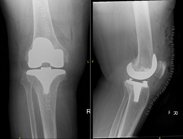
Total Knee Replacement
All three compartments are resurfaced: the femoral condyles and the tibial plateau are replaced with metal components, a medical-grade polyethylene spacer sits between them, and the patella is resurfaced where needed. Robotic guidance allows sub-millimetre implant positioning accuracy.
- Suitable for multi-compartment or whole-joint arthritis
- Robotic-assisted (MAKO, ROSA, CORI) for precise alignment
- Modern implants designed for a 20 to 25 year lifespan
- Most patients mobilising the same day as surgery
- Day-case surgery available for suitable patients
Partial Knee Replacement
Only the affected compartment is replaced, typically the medial (inner) side. The healthy cartilage, bone, and both cruciate ligaments in the other compartments are left completely intact, giving a more natural-feeling knee after recovery.
- Suitable for single-compartment arthritis with intact ligaments
- Faster recovery and less blood loss than total replacement
- More natural knee kinematics and proprioception
- Not suitable for inflammatory arthritis or BMI over 40
- Revision to total replacement possible if other compartments later deteriorate
Mr Hussain performs knee replacement surgery at the Royal Orthopaedic Hospital Birmingham. Explore the full range of knee replacement options in Birmingham, including total, partial, and robotic-assisted surgery.
How successful is knee replacement for arthritis?
Knee replacement is one of the most effective procedures in orthopaedic surgery. National Joint Registry data shows approximately 90 per cent of total knee replacements are still functioning at 15 years. Patient satisfaction after knee replacement is approximately 80 to 85 per cent, and robotic-assisted surgery is improving implant positioning accuracy, which is expected to further improve long-term outcomes.
Knee replacement outcomes have improved steadily over recent decades, driven by advances in implant design, surgical technique, robotic-assisted positioning, and accelerated recovery protocols. The NJR's 22nd Annual Report (2025) confirmed continued year-on-year reductions in revision rates across England, Wales, Northern Ireland, and the Isle of Man.
Robotic-assisted surgery, in which Mr Hussain holds certifications on three platforms (MAKO, ROSA, and CORI), allows the surgical plan to be personalised to each patient's anatomy before the first incision, and then executed with sub-millimetre accuracy during the procedure. The goal is a well-aligned, well-balanced knee that functions naturally and lasts as long as possible.
Expertise in knee arthritis treatment in Birmingham
Consultant at the Royal Orthopaedic Hospital
Mr Hussain practises at the Royal Orthopaedic Hospital Birmingham, one of the largest specialist orthopaedic hospitals in Europe, alongside Priory Hospital Edgbaston and Harborne Hospital.
3,000+ arthroplasty cases
From a total of more than 5,000 procedures performed, giving the operative volume and case complexity required for consistently excellent outcomes. Read more about Mr Hussain's training and background.
MAKO, ROSA, and CORI robotic certifications
Mr Hussain holds certifications on all three major robotic knee replacement platforms, allowing robotic-assisted surgery to be offered to all suitable patients regardless of which platform is available at their chosen hospital.
Complex revision surgery expertise
Mr Hussain performs revision knee replacement for patients whose primary replacement has failed, including for infection, loosening, and instability. See revision knee surgery for more information.
Doctify Outstanding Patient Experience 2024, 2025, and 2026
Awarded in three consecutive years, recognising consistently high patient-reported outcomes and communication throughout the surgical journey.
4.98 out of 5 from verified reviews on Doctify. Outstanding Patient Experience Award 2024, 2025, and 2026.
Frequently asked questions about knee arthritis
For more questions about surgery, recovery, fees, and what to expect, see the full frequently asked questions page or read recent patient testimonials.