Hip Fracture (Broken Hip)
A hip fracture is a break in the upper end of the femur, most commonly the femoral neck or the intertrochanteric region. Surgery is almost always required, with the operation chosen according to the fracture type and patient fitness. Mr Shakir Hussain, Consultant Orthopaedic Surgeon at the Royal Orthopaedic Hospital Birmingham, performs total hip replacement and partial hip replacement (hemiarthroplasty) for hip fracture and manages failed prior fixation and post-traumatic arthritis.
What is a hip fracture?
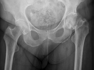
A hip fracture is a break in the upper end of the femur (thigh bone), close to the hip joint. The break sits either inside the joint capsule (intracapsular, typically a femoral neck fracture) or outside it (extracapsular, typically an intertrochanteric fracture). The distinction matters because intracapsular fractures disrupt the femoral head's blood supply, while extracapsular fractures preserve it. This single anatomical fact drives almost every treatment decision that follows.
The UK records approximately 79,000 hip fractures every year, according to the 2024 National Hip Fracture Database (NHFD). The average patient is 83 years old, around 70 per cent are women, and the dominant mechanism is a low-energy fall from standing height onto a bone weakened by osteoporosis. Younger patients suffer hip fractures only through high-energy trauma such as road traffic collisions or falls from height.
The two main anatomical types are:
- Intracapsular fractures (femoral neck). The break sits above the intertrochanteric line, within the joint capsule. The small arteries that feed the femoral head run up the neck and are torn or compressed by the fracture. A displaced intracapsular fracture risks avascular necrosis and non-union if simply fixed, so the femoral head is usually replaced.
- Extracapsular fractures (intertrochanteric, subtrochanteric). The break lies outside the capsule, through cancellous bone with a preserved blood supply. These fractures heal reliably once stabilised, so they are treated with internal fixation rather than replacement.

How does a hip fracture present?
The classic presentation is sudden severe groin or hip pain after a low-energy fall in an older person, with inability to bear weight and a leg that lies shortened and externally rotated. Around 3 per cent of hip fractures are radiographically occult: the patient may have only mild groin pain on movement and can occasionally still weight-bear. Persistent hip pain after any fall in an older patient should be investigated, even when the initial X-ray looks normal.
The textbook signs of a displaced hip fracture are easy to recognise, but subtler fractures (impacted femoral neck, stress fractures) can mislead families and even some clinicians. Mr Hussain sees both ends of the spectrum.
Sudden severe groin or hip pain after a fall
The pain comes on instantly with the fall. Patients describe it as a sharp, sickening pain in the groin or front of the hip, often with an audible crack.
Inability to bear weight
The patient cannot stand or walk on the affected leg after the fall. Attempting to do so causes severe pain. This is the cardinal sign of a displaced fracture.
Shortened, externally rotated leg
The leg lies shorter than the other side and is rotated outward, with the foot pointing away. This is caused by the muscle pull around the broken femur.
Bruising and swelling over the hip
Often subtle initially, developing over hours to days as blood from the fracture spreads through the soft tissues.
Occult fracture with only mild pain
About 3 per cent of fractures, particularly impacted femoral neck fractures, present with only mild groin pain on weight-bearing. Persistent pain after a fall warrants MRI even if X-rays appear normal.
What to do if you suspect a hip fracture
Do not try to walk on the affected leg. Call 999 or get to an A&E department for an X-ray. Hip fractures are time-critical: surgery within 36 hours is the strongest predictor of good recovery.
What causes a hip fracture?
The dominant cause of hip fracture in older patients is a low-energy fall onto a bone weakened by osteoporosis. Risk rises sharply after age 75 and is two to three times higher in women than in men. Factors that increase falling (poor vision, polypharmacy, cognitive impairment, sarcopenia) and factors that weaken bone (osteoporosis, vitamin D deficiency, prior fragility fracture) both contribute.
Hip fracture in older patients is fundamentally a disease of bone fragility plus falls. Either alone would be tolerable; together they produce the 79,000 annual UK fractures recorded by the NHFD. Younger patients suffer hip fractures only through high-energy trauma.
The most important risk factors are:
- Age over 75. The single strongest demographic risk factor, reflecting age-related bone loss and increasing falls.
- Osteoporosis. Bone mineral density two or more standard deviations below the young adult mean. The cornerstone risk factor for fragility fracture.
- Prior fragility fracture. A previous wrist, vertebral, or hip fracture roughly doubles the risk of a future hip fracture.
- Female sex. Women are affected approximately two to three times more often than men, particularly after the menopause.
- Vitamin D deficiency. Common in the UK, particularly in housebound patients. Contributes to both bone weakness and falls.
- Sarcopenia and frailty. Muscle loss reduces protection against falls and the impact of a fall on the hip.
- Cognitive impairment. Dementia substantially increases fall risk and complicates recovery.
- Polypharmacy. Particularly benzodiazepines, Z-drugs (zopiclone, zolpidem), antipsychotics, antidepressants, sedating antihistamines, and antihypertensives that cause postural hypotension.
- Visual impairment and environmental hazards. Poor lighting, loose rugs, and untreated cataracts are all modifiable.
In younger patients (under 60), hip fractures generally require high-energy trauma such as a road traffic collision, fall from height, or sports injury, and warrant a different work-up to exclude underlying bone disease.
How is a hip fracture diagnosed?
Standard AP and lateral hip X-rays diagnose most hip fractures immediately. Per NICE NG124, an MRI within 24 hours is the next investigation if clinical suspicion remains despite normal X-rays. T1-weighted MRI approaches 100 per cent sensitivity for occult femoral neck fractures. CT is acceptable if MRI is contraindicated or unavailable in the 24-hour window.
The vast majority of hip fractures are diagnosed in A&E within an hour of arrival. The clinical pathway is well established and time-critical: per BOAST and NICE NG124, surgery should happen on the day of admission or the day after, and within 36 hours where the patient is medically optimised.
Investigations used in hip fracture:
- AP and lateral hip X-rays. First-line imaging. Most hip fractures are diagnosed at this point, with the type (intracapsular versus extracapsular) and degree of displacement informing the surgical plan.
- MRI within 24 hours. Indicated when clinical suspicion remains despite normal X-rays, particularly for patients who cannot weight-bear after a fall. T1-weighted MRI is the most sensitive test for occult femoral neck fracture.
- CT. An acceptable alternative when MRI is contraindicated (pacemaker, severe claustrophobia) or not available within the 24-hour window. Also used for complex fracture planning.
- Bloods and pre-operative workup. Full blood count, urea and electrolytes, coagulation, group and save, ECG, chest X-ray, and an anaesthetic and orthogeriatric review to identify and treat dehydration, anaemia, electrolyte derangement, and cardiac disease before surgery.
- Cognitive screen. The 4AT delirium and cognition screen is administered on admission to detect existing cognitive impairment and risk of postoperative delirium.
What happens after a hip fracture?
Hip fractures are time-critical injuries that require admission to hospital and surgery within 36 hours wherever possible. Most patients are managed acutely by the on-call NHS orthopaedic team using a coordinated pathway involving orthogeriatric review, anaesthetic optimisation, prompt surgery, day-one mobilisation, and Fracture Liaison Service follow-up to prevent the next fracture.
The UK NHS hip fracture pathway is delivered through more than 175 hospitals and audited by the National Hip Fracture Database (NHFD). Performance is measured against Key Performance Indicators including:
- Prompt surgery. Operation on the day of, or day after, admission. Delays beyond 36 hours are associated with worse outcomes.
- Orthogeriatric review. A specialist physician for older people reviews every hip fracture patient to manage medical comorbidities, medication, and delirium risk.
- Multidisciplinary pre-operative assessment. Anaesthesia, surgery, orthogeriatrics, physiotherapy, and pharmacy plan the operation together.
- Day-one mobilisation. Physiotherapy gets the patient sitting out of bed the day after surgery and walking with support shortly after.
- Delirium screening. The 4AT score is repeated regularly to detect and treat postoperative delirium, which delays recovery if missed.
- Bone-health follow-up. Referral to a Fracture Liaison Service for DEXA scan and osteoporosis treatment to reduce the risk of a future fracture.
This pathway is delivered free at the point of use through the NHS. There is no private alternative for the acute admission. Private consultation with Mr Hussain becomes relevant for problems that develop after the acute episode (failed fixation, non-union, post-traumatic arthritis), for second opinions on implant choice or rehabilitation, or for planning ahead in osteoporotic patients who want to reduce their risk of fracture.
If you take prescribed medication, particularly blood-thinners or osteoporosis drugs, please review Mr Hussain's patient guide on medications to pause before hip or knee surgery.
How is the surgical choice made?
The choice between total hip replacement, partial hip replacement (hemiarthroplasty), and internal fixation is dictated primarily by the fracture type. For displaced femoral neck fractures in fit, mobile, cognitively well patients, NICE NG124 (updated 2023) recommends total hip replacement. For other displaced femoral neck fractures, cemented hemiarthroplasty is the standard. For extracapsular fractures, internal fixation (sliding hip screw or intramedullary nail) is the right choice.
The NICE NG124 (2023 update) framework drives this decision in UK practice. The criteria for offering total hip replacement rather than hemiarthroplasty in displaced femoral neck fracture are:
- Able to walk independently outdoors with no more than the use of a stick before the fracture
- Not cognitively impaired
- Medically fit for anaesthesia and the procedure
- Expected to remain independent in activities of daily living beyond two years
Patients who meet all four criteria are offered total hip replacement, which has lower long-term revision rates and better function than hemiarthroplasty in this group. Patients who do not meet the criteria are offered cemented hemiarthroplasty, which is faster, less invasive, and dislocates less often.
For extracapsular fractures (intertrochanteric and subtrochanteric), the surgical choice is between fixation devices rather than replacement: a sliding hip screw or a cephalomedullary intramedullary nail. The blood supply to the femoral head is intact in these fractures, so the bone can heal once stabilised.
Private consultations with Mr Hussain are available at the Royal Orthopaedic Hospital, Priory Hospital Edgbaston, and Harborne Hospital. See the private consultation and surgery fees page for pricing and recognised insurers, or book a consultation directly to discuss failed prior fixation, post-traumatic arthritis, or a second opinion before an elective procedure.
What are the surgical options for hip fracture?
Three operations cover almost every hip fracture: total hip replacement for fit, mobile patients with a displaced femoral neck fracture; cemented hemiarthroplasty (partial hip replacement) for other displaced femoral neck fractures; and internal fixation with a sliding hip screw or intramedullary nail for extracapsular fractures and undisplaced femoral neck fractures. Hip resurfacing is not used for acute hip fracture in any current UK guideline.
Each option matches a fracture type and a patient profile. Mr Hussain performs total hip replacement and hemiarthroplasty for fracture, and treats the late consequences (failed fixation, post-traumatic arthritis) of all three.
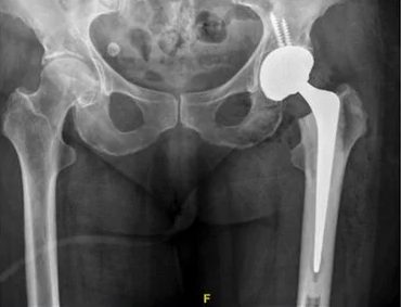
Total Hip Replacement
The femoral head and the acetabular surface are both replaced. NICE NG124 (2023) recommends THR for displaced femoral neck fractures in patients who walked independently outdoors with no more than a stick, are cognitively well, and are medically fit. Cemented femoral fixation is recommended in this fragility population.
- Best long-term function and lowest revision rate for fit fracture patients
- Cemented stem fixation per NICE NG124
- Restores anatomic geometry and leg length
- Same components used in elective hip replacement for arthritis
Hemiarthroplasty (Partial Hip Replacement)
Only the femoral head is replaced; the patient's own acetabulum is retained. Used for displaced femoral neck fractures in patients who do not meet the NICE NG124 criteria for total hip replacement. NICE 2023 advises hospitals to standardise on one cemented femoral component for this operation.
- Faster operation, less blood loss, lower dislocation rate
- Cemented polished tapered or anatomical stem
- Suitable for patients with limited pre-fracture mobility
- Acceptable when life expectancy is shorter than the projected implant survival
For extracapsular (intertrochanteric and subtrochanteric) fractures, fixation rather than replacement is the right operation. The blood supply to the femoral head is intact, so the bone can heal once stabilised with a sliding hip screw or an intramedullary nail. Hip resurfacing is not used for acute hip fracture: a displaced intracapsular fracture disrupts the blood supply to the femoral head, which would lead to avascular necrosis under a resurfacing cap.
For a deeper comparison of the elective hip replacement options, see Mr Hussain's patient guide on hip resurfacing versus total hip replacement (relevant to elective arthritis surgery, not acute fracture).
What are the outcomes after hip fracture surgery?
Modern hip fracture care has dramatically improved outcomes. The 2024 National Hip Fracture Database reports 30-day mortality below 7 per cent in the audited population, with around half of patients regaining their pre-fracture mobility by 120 days. The strongest predictors of good recovery are surgery within 36 hours, orthogeriatric review, day-one mobilisation, and early bone-health follow-up.
Hip fracture mortality has fallen substantially over the last two decades through coordinated multidisciplinary care. The NHFD captures more than 70,000 hip fractures every year and publishes benchmarked unit-level performance, driving continuous improvement across the 175-plus hospitals contributing data. The figures below reflect the 2024 annual report on 2023 data.
Expertise in hip fracture and post-fracture surgery in Birmingham
Consultant at the Royal Orthopaedic Hospital
Mr Hussain practises at the Royal Orthopaedic Hospital Birmingham, one of the largest specialist orthopaedic hospitals in Europe, alongside Priory Hospital Edgbaston and Harborne Hospital.
3,000+ arthroplasty cases
From a total of more than 5,000 procedures performed, giving the operative volume and case complexity required for consistently excellent outcomes. Read more about Mr Hussain's training and background.
British Hip Society Travelling Fellowship
Trained at ENDO-Klinik Hamburg under Professor Thorsten Gehrke and Professor Mustafa Citak, the international reference centre for complex hip surgery.
Both hip replacement and hip resurfacing
Many surgeons offer only one technique. Mr Hussain trained in both, allowing the choice to be tailored to the individual patient rather than to a single technique.
Doctify Outstanding Patient Experience 2024, 2025, and 2026
Awarded in three consecutive years, recognising consistently high patient-reported outcomes and communication.
4.98 out of 5 from verified reviews on Doctify. Outstanding Patient Experience Award 2024, 2025, and 2026.
Frequently asked questions about hip fracture
For more questions about surgery, recovery, fees, and what to expect, see the full frequently asked questions page or read recent patient testimonials.